Supracondylar Amputation: Definition And Intervention
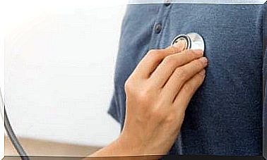
The aim of a supracondylar amputation is that the rest of the body heals well and without complications, so that the patient can return to everyday life in the foreseeable future. It is also important to offer him as much quality of life as possible.
The aim of a supracondylar amputation is that the rest of the body part heals well and without complications, so that the patient can return to everyday life in the foreseeable future. It is also important to offer him as much quality of life as possible.
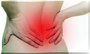
This surgical procedure involves removing a limb above the knee condyles. Between 50 and 60 percent of non-traumatic amputations of this type are due to diabetes.
A supracondylar amputation is applied when the previous treatment alternatives have failed. Therefore, acceptance must be achieved on the part of the patient and a clear goal formulated for him: to protect his health and guarantee a high quality of life.
The resulting stump should heal well and be stable so that a prosthesis can be put on as quickly as possible. This makes it possible for the person affected to lead a largely normal life.
Supracondylar amputation: general principles
In general, a distinction can be made between smaller and larger amputations. Supracondylar amputation is one of the latter because it affects a larger area. However, every procedure, regardless of its severity, is a complex procedure that is based on a few basic aspects. These include the following:
- Treatment with antibiotics is always necessary. This is used to control infections or as a prophylactic measure.
- The hemostasis or the control of bleeding must be consistent. If a hematoma appears, it is a sign of necrosis or infection.
- There must be no tension at the edges of the skin. To prevent this from happening, it is necessary to be careful with the soft tissue.
- There should be an appropriate relationship between the bone section and the length of the skin and muscle tendons. This avoids tension and ensures good coverage of the bone.
- A traction of the nerve tracts should be carried out to prevent possible neuromas of the scar. This also applies to the articular cartilage and tendons.
- Avoid leaving bone fragments in the wound or forming a bulge.
- The surgical wound must be washed repeatedly with physiological or antiseptic serum before it is closed.
Indications and contraindications
The supracondylar amputation is used when healing from a previously performed infracondylar amputation has not promised success. Another reason is potential calf muscle spasms affecting the flexion of the knee joint.
The knee joint is lost in supracondylar amputation. To avoid complications with the prosthesis that the patient will be wearing, it is important that the residual limb is of suitable length.
This procedure is not recommended in the case of gangrene or an infection of the thigh.
method
The following steps outline how a supracondylar amputation is performed:
- The patient is on his back.
- The surgeon marks the incision site in the shape of a fish’s mouth, also called a shark’s mouth.
- Doctors make the incision with a cold scalpel.
- The subcutaneous tissue is cut open to the tendon plate (aponeurosis) or membrane that covers the muscles and is attached to the bone. An electric scalpel is used for this. In the case of a deep incision, care must be taken to ensure that sufficient tissue remains for the stump.
- Then the superficial and deep femoral vascular package as well as the sciatic nerve are identified, tied off and severed. It is necessary to use infiltration anesthesia.
- The thighbone is enclosed in its entire circumference.
- I m connection separate doctors the tissue that is connected to the thigh bone. The periosteum is used for this.
Then the following steps are necessary:
- The so-called Percy retractor is adapted in order to be able to amputate the femur. This instrument allows enough soft tissue to cover the stump.
- Then the bone is sawn through with the Gigli saw. For this, a 90 degree angle is created between the two ends of these. During this time, the area should be continuously cleaned with a physiological serum.
- Then the edges of the bone must be filed.
- The next step is to apply bone wax to the cut area. This is pressed onto the surface so that it sticks. The excess is disposed of.
- The stump should now be closed with synthetic, non-absorbable surgical suture material (Prolene). First, the surgeon closes the deepest muscle groups so that the bone surface is covered. Afterwards he sews the upper tendon plate.
- The tissue under the skin is closed with an absorbable, synthetic suture, better known as vicryl.
- The skin itself is sewn with a silk seam using the so-called mattress stitch.