Vascular Perforation Through Central Venous Catheter
Vascular perforation is a very rare complication. Only 10% of perforations occur in central veins. However, the correct diagnosis is usually made late, which represents an increased risk and can be fatal.
Vascular perforation occurs when a venous catheter is not placed correctly and the tip moves. This can happen at any time, regardless of where the catheter is located.
However, it is a very rare complication. Only 10% of perforations occur in central veins. However, the correct diagnosis is often made very late, which is an additional hazard and can be fatal.
Ultrasound is used to assess whether a particular vein is suitable for catheter placement . This enables the radiologist to plan a correct intervention. Learn more about different venous catheters and possible complications today .
Venous catheters: what types are there?

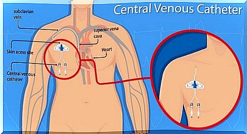
A venous catheter (indwelling venous cannula) is a thin plastic tube that is fed through the venous system into the upper or lower vena cava in front of the right atrium of the heart. The size, length and number of channels in the catheter are variable. They depend on the cause of use. Then we will describe the most frequently used venous catheters in more detail:
Peripheral venous catheter
It is a long catheter that is inserted through a peripheral vein on the arm or leg until it reaches the superior or inferior vena cava in front of the right atrium of the heart. One also speaks of an indwelling venous cannula or a peripheral venous access. This type of catheter is often used to give intravenous drugs or IV fluids.
Central venous catheter not tunneled under
A venous catheter that is not tunneled under can be larger and is therefore used for relatively large veins. These are veins that are more central, such as the jugular vein or the femoral vein.
Under-tunneled venous catheter
With a tunneled venous catheter, the entry into the vein is located within the subcutaneous tissue. Access is always possible, no need to change the needle. There are different sizes and types of tunneled catheters. Particularly noteworthy in this case are the safety and easy access to the cannula.
Port catheter
It is a system that allows permanent access to the venous or arterial blood circulation. The port catheter consists of one (or more) chambers and a tube.
Risk of vascular perforation and other complications from a central venous catheter
Serious complications can occur if the venous catheter is improperly placed. One possible consequence is vascular perforation. Therefore, the correct position must always be monitored.
A computed tomography with contrast is used to diagnose a vascular perforation. There are two types of risk with venous catheters: complications from improper insertion of the cannula and complications that occur in the body after insertion.
The most common risks include:
- Infection at the puncture site shortly after the catheter is placed
- Bleeding and bruises
- Pneumothorax: It is a pathological accumulation of air in the chest, which can lead to a collapse of a lung. This in turn causes the lungs to function poorly.
- In rare cases, an arterial vessel is incorrectly positioned instead of the catheter being placed in the vein. If this is the case, the doctor must remove the catheter again. In most cases, the artery will heal on its own, but surgery may sometimes be necessary.
Late infection through a venous catheter
Two types of late infection can occur with a venous catheter: infection of the skin at the puncture site or infection in the bloodstream.
If the catheter does not adhere well to the skin, there is a risk that it may accidentally come off. If this is the case, you have to call a doctor as soon as possible. In the meantime, pressure is applied to the puncture site with a sterile wound pad.
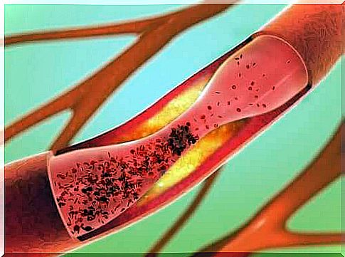
A blood clot could clog the venous catheter, regardless of the type. The doctor can then use a blood thinner to dissolve the clot, but in some cases they may have to remove the catheter.
Only rarely does the patient experience irregular heartbeats, which is also related to the catheter.